Menu

 Edwin Y. Endo, OD Optometrists, Eye Doctors Of Honolulu

Leading Provider in Professional Optometry Eye Care and highly regarded establishment offering state of the art Practice & progressive Vision eye exams with Excellence. The Art of Caring.

header photo

Glaucoma Facts and Statistics

  • After cataracts, glaucoma is the leading cause of blindness among African Americans.
  • Estimates put the total number of suspected cases of glaucoma at over 60 million worldwide.
  • Blindness from glaucoma is 6 to 8 times more common in African Americans than Caucasians.
  • African Americans are 15 times more likely to be visually impaired from glaucoma than Caucasians.
  • It is estimated that over 2.2 million Americans have glaucoma but only half of those know they have it.
  • In the U.S., more than 120,000 are blind from glaucoma, accounting for 9% to 12% of all cases of blindness.
  • Glaucoma is the second leading cause of blindness in the world, according to the World Health Organization.
  • The most common form, open-angle glaucoma, accounts for 19% of all blindness among African Americans compared to 6% in Caucasians.
  • Other high-risk groups include: people over 60, family members of those already diagnosed, diabetics, and people who are severely nearsighted.
  • Glaucoma causes irreversible blindness and many (50%) of the affected people are unaware of their condition.
  • Glaucoma is second only to cataract as the leading cause of preventable blindness in the world. It is estimated that over 65 million people throughout the world are affected by glaucoma.
  • Corticosteroid drugs used to treat eye inflammations and other diseases can trigger glaucoma in some people. Treatments include, but are not limited to, medicines, laser surgery, or conventional surgery.

As far as illnesses go, a diagnosis of glaucoma can be particularly devastating. The disease is sneaky: It develops slowly, often without warning, and can lead to irreversible blindness if it’s not treated in time. Glaucoma is caused by fluid building up in the front part of your eye, increasing pressure and damaging the optic nerve — a kind of electric cable that sends visual information from the eyeball to the brain.

More than 2.7 million Americans over 40 have glaucoma, but only half are aware of it, according to the American Academy of Ophthalmology. The disease is one of the world’s leading cause of blindness. Particularly at risk: People over the age of 40. The Glaucoma Research Foundation reports that you’re six times more likely to get glaucoma if you’re over 60. Also more vulnerable than average are those with a family history of the disease, African Americans, Hispanics, people with diabetes and smokers.


Glaucoma infographic
 

Classification of Glaucoma

Glaucoma is classified as primary (due to a primary abnormality in the aqueous drainage pathways) or secondary (to other ocular disease).

Primary Glaucoma

Primary glaucoma is rare in the cat, but common in the dog. It is divided into those forms where there is a preceding gross abnormality in the development of the iridocorneal angle that can be detected by gonioscopy and those where the opening into the ciliary cleft (iridocorneal angle) appears normal.

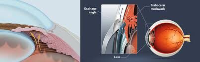
 Narrow angle and goniodysgenesis. These are the commonest forms of primary glaucoma in dogs. The drainage angle is abnormally formed. In narrow angle there is an abnormally narrow opening into the ciliary cleft. With goniodysgenesis the opening into the ciliary cleft is spanned by an abnormally differentiated pectinate ligament. The malformation of the drainage apparatus is heritable and the more severely affected animals will develop glaucoma. Other, poorly understood factors also play a role because although these abnormalities are present after ocular maturation, glaucoma does not develop until later in age (often middle age). There is a suggestion that further narrowing of the angle may occur with age. Screening dogs of at risk breeds can be performed to identify individuals with the predisposing anatomical abnormalities. Many breeds of dog are affected including Cocker spaniel, the Basset hound, arctic circle breeds (Samoyed, Siberian husky, Norwegian elkhound), Bouvier, terrier breeds, miniature poodle, Chow chow, Shar pei. This form of glaucoma typically has a very acute onset and results in very marked increases in IOP (above 60mmHg is common).

 Open angle glaucoma. This is the commonest form of glaucoma in man but is uncommon in dogs. It typically has a slow and insidious onset with globe enlargement and accompanying changes (splits in Descemet's membrane of the cornea and lens subluxation) as the first outward sign of the disease. Initially the IOP is only slightly to moderately elevated. As the disease progresses more severe elevations of IOP with accompanying pain may develop.

Secondary Glaucoma

Secondary glaucoma occurs as a complication of, or sequel to, other ocular disease or injury. Any disease process can interfere with aqueous outflow and result in secondary glaucoma. Causes include:

 Primary lens luxation. Anterior luxation of the lens accompanied by vitreous prolapse leads to pupil block and glaucoma. Typically acute-onset, require emergency medical reduction in IOP followed by lens extraction.

 Iridocyclitis. Chronic anterior uveal inflammation can lead to secondary glaucoma by either extensive posterior synechiae (iris to lens adhesions) and pupil block, or impairment of aqueous passage through the drainage apparatus.

 Trauma

 Hyphema. Bleeding into the anterior chamber (particularly if recurrent) can lead to impairment of aqueous drainage and secondary glaucoma.

 Retinal detachment. Can lead to development of a pre-iridal fibrovascular membrane that can obstruct the drainage angle.

 Neoplasia. Infiltrating the drainage angle.

 Ocular melanosis. This condition is a hereditary condition in cairn terriers. Proliferation of melanocytes within the eye eventually leads to impairment of aqueous drainage. Glaucoma has an insidious onset and leads to globe enlargement prior to eventual loss of vision. It is difficult to control.

Clinical Signs of Glaucoma

The clinical signs of glaucoma vary considerably depending on the etiology, the duration and the extent of pressure rise.

 Acute glaucoma with grossly elevated IOP--typical signs with primary narrow angle or goniodysgenesis glaucoma or secondary to lens luxation include:

 Severe pain (blepharospasm, photophobia, enophthalmos, elevation of the nictitating membrane, diffuse facial pain--trigeminal neuralgia--winces if head touched), slight epiphora. Altered behavior (hiding, off food). The signs of severe pain reduce a few days after onset.

 Episcleral and conjunctival vascular injection ("Red-Eye") occurs due to blockage of normal venous drainage and engorgement of anterior ciliary veins.

 Corneal edema (reversible) due to increased hydrostatic pressure which exceeds the endothelium's capacity to pump fluid out of the stroma.

 Pupillary dilation--Dilation or unresponsiveness to light stimulation doesn't occur until the IOP is about 40 mmHg or higher. Pupil is typically mid-dilated in the acute narrow angle/goniodysgenesis glaucomas. May be constricted in some secondary glaucomas

 Loss of vision As the pressure rises above the 40s vision is reduced and then lost--in most cases this is initially reversible with very prompt emergency treatment.

 Early stages of glaucoma with only moderately elevated IOP--open angle glaucoma and many secondary glaucomas fall into this category. At this stage pain is not an obvious feature and vision loss is not yet significant. There may be a mild episcleral injection. The most obvious sign will be globe enlargement. However this will not be apparent until the IOP has been elevated for some time.

 Chronic glaucoma--Primary open angle glaucoma and some secondary glaucomas may first present when they already have some chronic changes.

 Buphthalmos--globe enlargement; occurs more quickly in young animals. If this has developed in an eye with narrow angle glaucoma or goniodysgenesis the eye will probably be blind. Young animals and those with primary open angle glaucoma and glaucoma due to ocular melanosis may still have useful vision at the time when buphthalmos has developed.

 Corneal changes--within a few days of development of severe IOP rises deep corneal vascularization may develop. More chronic changes include: exposure keratitis (may occur if the animal is unable to blink over the buphthalmic globe); corneal (Haab's) striae--gray, linear streaks in the cornea due to breaks in Descemet's membrane.

 Scleral thinning

 Decreased evidence of pain--but condition remains painful

 Episcleral vascular engorgement

 Subluxated/luxated lens--zonular breakdown, vitreal degeneration with loss of support for lens; subsequent cataract development

 Iris degeneration

 Ciliary body degeneration (can eventually result in phthisis bulbi)

 Cataract

 Funduscopic lesions

 Optic atrophy & cupped optic disc--posterior bowing of the lamina cribrosa

 Retinal degeneration--thinning of the retina, increased tapetal reflectivity and retinal vascular attenuation

Although the damage from glaucoma cannot be reversed or restored, early diagnosis can help prevent further damage by reducing eye pressure. By following the treatment plan and keeping up with regular eye exams, you can help mitigate and control the damage and effect glaucoma has in your life. Call us today and avoid further eye damage from glaucoma!

Definitions & Additional Information

Glaucoma: An optic neuropathy associated with progressive death of retinal ganglion cells and their axons, and associated visual field loss. The characteristic changes of the optic nerve head that distinguish glaucoma from other optic neuropathies include excavation and undermining of the neural and connective tissues.

Primary open-angle glaucoma (also chronic open-angle glaucoma): Glaucoma in the setting of an eye with a visibly open anterior chamber angle (between the iris and anterior sclera/peripheral cornea) and no other ocular or systemic disorder that might result in glaucoma. 

Secondary open-angle glaucoma: Glaucoma in the setting of an eye with a visibly open anterior chamber angle (between the iris and anterior sclera/peripheral cornea) and some other ocular or systemic disorder that can result in glaucoma. Examples of secondary open-angle glaucomas include pigment dispersion syndrome, pseudoexfoliation syndrome, and steroid-induced glaucoma.

Glaucoma suspect: A nonspecific term describing someone at higher than average risk of having or developing glaucoma. In the case of open-angle glaucoma, this risk may be increased due to elevated intraocular pressure (ocular hypertension), an optic nerve with an appearance consistent with the structural changes caused by glaucoma, a significant family history of the disease, or a racial background known to confer higher rates of glaucoma. It is currently possible to estimate the risk of future glaucoma only in some patients in the ocular hypertensive group.

Treatments for Open-Angle Glaucoma

Medical, laser, and incisional surgical treatments are used to treat glaucoma. The most common currently used medical treatment includes several classes of eye drops, including prostaglandin analogs, beta-adrenergic antagonists, oral and topical carbonic anhydrase inhibitors, and alpha-adrenergic agonists. Laser trabeculoplasty is an office-based procedure that lowers the IOP by increasing the outflow of aqueous humor from the eye. Incisional surgery to lower the IOP comprises procedures that have been performed for decades, such as trabeculectomy and aqueous drainage device surgery, as well as a host of newer procedures, such as nonpenetrating deep sclerectomy, canaloplasty, endoscopic cyclophotocoagulation, and alternative methods of trabecular bypass.

Definitions of Laser and Incisional treatments

Laser trabeculoplasty: A procedure in which laser energy (argon, YAG, diode) is applied to the trabecular meshwork in an effort to reduce the resistance to outflow for aqueous humor. The procedure is performed as part of an office visit and requires topical anesthesia and a mirrored contact lens.

Trabeculectomy: The most commonly performed incisional surgery for lowering intraocular pressure in glaucoma patients. Under local anesthesia, a passageway is created at the limbus (junction between the cornea and sclera) that allows the aqueous humor to flow from the anterior chamber to the space between the sclera and the conjunctiva, thereby lowering the intraocular pressure. The hallmark of a trabeculectomy is the fluid-filled bleb (blister) present on the surface of the eye underneath the upper eyelid.

Trabeculotomy: An incisional surgery procedure generally used to lower intraocular pressure in glaucoma affecting infants and children. A metal probe or a suture is passed into Schlemm’s canal, a structure into which aqueous humor passes as it exits the eye. The probe is used to disrupt tissue that is typically impeding outflow of aqueous humor from the eye, thereby increasing outflow and decreasing the intraocular pressure. Some surgeons also use trabeculotomy in the treatment of glaucoma in adults.

Aqueous drainage devices: Any of a number of plastic implants used in the surgical management of glaucoma with the aim of lowering the intraocular pressure. All devices consist of a tube that is inserted into the eye and a plate connected to the tube that is sewn to the sclera and covered by conjunctiva. Aqueous humor moves through the tube and out of the eye to drain on top of the plate into the space between the plate and the conjunctiva.

Cyclophotocoagulation: A procedure in which laser energy is used to damage the ciliary processes, reducing the amount of aqueous humor that they produce and thereby lowering the intraocular pressure. The procedure can be performed through the sclera (external cyclophotocoagulation) or from the inside of the eye (endocyclophotocoagulation).

Deep sclerectomy: A procedure in which the surgeon makes an opening in the conjunctiva to expose the sclera. The surgeon dissects a partial-thickness flap about 5 mm in width to about one-third depth in the sclera at the limbus. A second flap is dissected below this flap in order to leave a very thin layer of tissue and to expose Schlemm's canal. This underlying flap of scleral tissue is removed, and the surgeon grasps the roof of Schlemm's canal and removes a strip that is about 3 mm in length. Aqueous humor is able to permeate the remaining tissue without a full-thickness hole being necessary. The external flap is then sutured in its original position and the conjunctiva is sewn back in place.

Viscocanalostomy: A surgical procedure that is the same as for deep sclerectomy (see above) but also includes viscoelastic injected into Schlemm's canal in a circumferential fashion in an effort to dilate Schlemm's canal. The external flap is then sutured in its original position and the conjunctiva is sewn back in place.

Canaloplasty: A procedure that begins with a combined deep sclerectomy and viscocanalostomy procedure (see above), after which a microcatheter with an illuminated tip is passed through Schlemm's canal for 360 degrees. A 10-0 Prolene suture is tied to the catheter and threaded around Schlemm's canal for 360 degrees. The two ends of this suture are tied under tension in an effort to expand Schlemm's canal. The external flap is then sutured in its original position and the conjunctiva is put back in place.

Trabectome™: A procedure in which the surgeon makes a 1.7 mm incision through the peripheral cornea and injects viscoelastic into the anterior chamber. The Trabectome device is then introduced into the anterior chamber and, under visualization using direct gonioscopy with an operating microscope, the Trabectome is used to ablate about one quadrant of trabecular tissue. The Trabectome uses low-energy electrical pulses to vaporize the trabecular tissue, and aspiration is used to remove it. The viscoelastic is removed and the corneal wound is sutured closed.

iStent™: A device placed into Schlemm’s canal. The Glaukos Trabecular Micro-Bypass Stent (iStent) is made of nonferromagnetic titanium. One end sits in the anterior chamber and the posterior end sits in Schlemm’s canal, allowing fluid to bypass the trabecular meshwork. The device is inserted under direct visualization (using direct gonioscopy) through a 3 mm temporal clear corneal incision. After viscoelastic is placed in the anterior chamber, the applicator is passed through the incision and the device is anchored into Schlemm’s canal in the nasal angle. Viscoelastic is removed with irrigation and aspiration.

Gold shunt: A device that connects the anterior chamber to the suprachoroidal space. The SOLX™ Gold Shunt is a 24-karat gold rectangle (3.2 x 5.2 mm). There are two plates with grooves in them to allow flow from the higher pressure anterior chamber to the lower pressure suprachoroidal space. The conjunctiva is disinserted at the limbus, and a full-thickness scleral incision is created 2 mm posterior to the limbus. A crescent blade is used at 90 percent scleral depth to direct the anterior portion of the shunt to the anterior chamber and to cut posteriorly 2 to 3 mm to direct the posterior segment into the suprachoroidal space. The scleral incision is closed with 10-0 nylon sutures and the conjunctiva is closed.

Incisional Treatments for Glaucoma


Currently Available Methods to Treat Open Angle Glaucoma Series:

  1. Currently Available Methods to Treat Open Angle Glaucoma
  2. Beta-blockers, Selective Alpha Adrenergic Agonist, CAIs
  3. Prostaglandin Analogs, Cholinergic Receptors Agonists, Fixed Combination Agents
  4. Carbonic Anhydrase Inhibitors (CAIs)
  5. Laser Trabeculoplasty
  6. Continuous Wave and Micropulse® Cyclophotocoagulation
  7. Trabeculectomy and Glaucoma Drainage Devices
  8. Ab-Externo Canaloplasty
  9. Ab-Interno Canaloplasty
  10. Trabeculotomy
  11. IStent®, Cypass® Microstent, Xen® 45 Gel Stent, Cataract Surgery
  12. Next-Generation Glaucoma Medications and Surgeries
  13. iStent Supra®, Hydrus™ Microstent, and InnFocus MicroShunt®
  14. Canaloplasty with Stegmann Canal Expander


Selective Laser Trabeculoplasty vs Topical Medication as Initial Glaucoma Treatment

Management of Glaucoma

In deciding on the best approach to managing a case of glaucoma the cause of the glaucoma must be identified. For example glaucoma secondary to lens luxation requires a different approach to say glaucoma due to an intraocular tumor or a case of primary glaucoma.

Medical Management

Several classes of drug are available to lower intraocular pressure but not all are effective in all species. A combination of several drugs may be used.

Prostaglandin Analogues

e.g., latanoprost, travoprost and bimatoprost Cause a profound reduction in the production of aqueous. Use for emergency reduction in IOP in dogs, not effective in cats. For maintenance use q12-q24hr. Cause significant miosis which may exacerbate underlying uveitis--also may increase risk of pupil block.

Osmotic Diuretics

Emergency reduction in IOP--increase the osmotic gradient between plasma and the ocular cavities, dehydrating the vitreous. Less commonly used since prostaglandin analogues became available. e.g., Mannitol IV 20% 1-2 g/kg IV over 15-20 minutes. Glycerol, 50% oral solution 1 to 2 ml/kg.

Carbonic Anhydrase Inhibitors (CAIs)

Decrease aqueous humor production usually applied topically (e.g., dorzolamide, brinzolamide). Useful for longer-term control.

Beta Blockers

Timolol Betaxolol Levobunolol--reduce aqueous formation.

Miotics (Applied Topically)

Increase aqueous humor outflow. Direct (pilocarpine) and indirect (the cholinesterases such as: demecarium bromide, phospholine) acting parasympathomimetics.

Surgical Management

Cyclodestructive Procedures

 Cyclocryotherapy

 Laser therapy (Transcleral diode laser, endolaser)

Alternative Drainage Pathways

 Aqueous drainage implants

Other Surgical Options for Blind Painful Eyes

 Enucleation

 Evisceration with intrascleral prosthesis


Link 

  • In this study, 167 patients with primary open-angle or exfoliation glaucoma were randomized to receive either selective laser trabeculoplasty (SLT) or topical medication as a first-line treatment. The authors compared the effect of these treatments on quality of life and clinical outcomes and found no differences between groups in terms of glaucoma-specific quality of life.

Selective Laser Trabeculoplasty as the Primary Treatment for Open-Angle Glaucoma 

  1. This commentary discusses the use of selective laser trabeculectomy as a first-line treatment for treatment-naïve primary open-angle glaucoma and ocular hypertension in view of the results from LiGHT study. 
  2. Overall, selective laser trabeculectomy appears to be subjectively effective and relatively risky, which may not allow it to be generalizable to all patient populations.

– Raza Shah, MD


Phacoemulsification vs Phacotrabeculectomy in Primary Angle-Closure Glaucoma With Cataract

Phacoemulsification alone reduces IOP and the need for glaucoma drugs in PACG eyes with cataracts for up to 5 years. Phacotrabeculectomy was more effective in reducing IOP and glaucoma drugs than phacoemulsification alone but was associated with more postoperative complications, up to 5 years after surgery.


What’s on Your Radar?

A rundown of some promising ophthalmic therapeutics coming down the pike.

Eric Devore, OD 

Katherine M. Mastrota, MS, OS, FAAO

The medically minded optometrist looks for ways to expand his or her role when treating patients with age-related eye disease, glaucoma, postoperative pain, diabetic eye disease, and other conditions. It’s important that we practice to the extent of our licenses, which means writing prescriptions and, perhaps more important, knowing about the drugs both we and our MD counterparts are prescribing.

Keeping current with the most commonly prescribed ophthalmic drugs helps us be cognizant of and better able to identify adverse reactions or side effects our patients may be experiencing. It also allows us to explore other options when side effects surface or when one class of drug is contraindicated.

This article highlights some promising therapeutics in the pipeline for the treatment of ocular diseases, conditions, and symptoms.

AMD AND DIABETIC EYE DISEASE

Abicipar Pegol

Although the pathophysiology of diabetic retinopathy differs in many ways from that of age-related macular degeneration (AMD), treatment for diabetic macular edema and proliferative diabetic retinopathy includes suppression of the same stimulus that leads to retinal neovascularization: ischemia and subsequent upregulation of VEGF. Abicipar pegol (Allergan) is a designed ankyrin repeat protein, or DARPin, a therapeutic with high affinity for VEGF-A.1 This new anti-VEGF drug has shown efficacy similar or superior to that of ranibizumab (Lucentis, Genentech) injections in patients with wet AMD.2,3 Two identical global phase 3 studies (SEQUOIA and CEDAR) demonstrated the efficacy of a 12-week fixed dosing regimen of abicipar, with 50% fewer injections than ranibizumab, in the treatment of patients with neovascular AMD.4

GLAUCOMA

Omidenepag Isopropyl

A pivotal phase 3 US development program (SPECTRUM) investigating the use of omidenepag isopropyl 0.002% (DE-117, Santen) for the treatment of glaucoma or ocular hypertension (OHT) was initiated in the United States last year.5 This follows positive results from phase 1/2, 2, and 2b dosing studies demonstrating that 0.002% omidenepag is the most appropriate dose and that the investigational drug performed similarly to latanoprost in reducing IOP. Omidenepag, a selective agonist for the prostanoid receptor EP2, was found to be generally safe and well tolerated in the earlier studies. Common side effects of prostaglandin agonists (eg, iris and eyelid pigmentation, abnormal eyelash changes, deepening of upper eyelid sulcus) were not observed during long-term (12 months) use in a Japanese study.5

Microdose Latanoprost Formulation

A proprietary microdose formulation of latanoprost (MicroProst, Eyenovia) is being developed as a potential first-line treatment for the reduction of IOP in patients with chronic angle-closure glaucoma, OHT, or primary open-angle glaucoma. In a phase 2 feasibility dose-finding study, 30 healthy volunteers received single 8-µL microdoses of 0.005% lantanoprost (0.4 µg) using a high-precision, piezo-print horizontal delivery system on 2 successive days. This treatment reduced diurnal IOP from baseline at 1 and 2 days after administration. Patients successfully self-administered the microdoses after training, and administration was well tolerated and did not result in adverse events.6 The company expects to enroll the first patient in a phase 3 trial in the first half of this year.

Sustained-Release Bimatoprost Implant

Bimatoprost SR (Allergan) is an intracameral bimatoprost implant designed for sustained release. A first phase 3 study of the formulation, completed in mid 2018, showed good results with the device over a 12-week period, with comparable efficacy to daily use of a prostaglandin analogue and superior efficacy to daily timolol.7 When we discuss with patients the option of initiating medical treatment or performing selective laser trabeculoplasty in the setting of primary open angle glaucoma, an intracameral implantable device may be a viable alternative to topical medications. In patients who opt for laser treatment first, if a desired endpoint is not reached, this implant, if approved, may be a reasonable next step before initiating lifelong topical medical therapy.

POSTOPERATIVE PAIN AND INFLAMMATION

Loteprednol Gel

Submicron loteprednol etabonate ophthalmic gel 0.38% (Bausch + Lomb) is an investigational formulation that uses novel submicron particles to facilitate ocular penetration of loteprednol into key anterior segment tissues (eg, iris, ciliary body, aqueous humor, and cornea). If approved, this ophthalmic gel would be the lowest concentration loteprednol corticosteroid formulation indicated for the treatment of postoperative inflammation and pain after ocular surgery.

In September, it was reported that this investigational formulation of loteprednol met dual primary efficacy endpoints in a clinical trial: It was significantly more effective than vehicle in completely resolving ocular inflammation and pain after cataract surgery.8 Additionally, submicron loteprednol etabonate ophthalmic gel 0.38% had an acceptable safety profile regardless of whether it was administered two or three times per day.

DRY EYE

Reproxalap

In a recently completed phase 2b clinical trial, reproxalap topical ophthalmic solution (Aldeyra Therapeutics) improved both signs and symptoms of dry eye. Aldehydes are posited to play a role in potentiating ocular surface inflammation through reactive aldehyde species (RASP). In patients with dry eye disease, RASP may contribute to ocular inflammation. By diminishing aldehyde levels, Aldeyra’s topical ocular aldehyde trap platform is a novel approach that may augment existing therapy, and, in severe cases, reduce or eliminate the need for corticosteroids. Additional indications for reproxalap may include treatment of uveitis, chronic allergic conjunctivitis, and atopic ocular disease.9 A phase 3 study assessing the use of reproxalap 0.25% and 0.50% in treating allergic conjunctivitis was completed in November; results are pending publication.

SkQ1

SkQ1 (Visomitin, Mitotech) is a small molecule described by Mitotech as a cardiolipin peroxidation inhibitor, a compound designed to reduce oxidative stress within mitochondria. The company is exploring its use for several indications, including treatment of moderate to severe dry eye disease. In a phase 2 US clinical study of 90 patients at a single center, the topical ophthalmic formulation demonstrated statistically significant superiority over placebo for several endpoints, including fluorescein staining, ocular discomfort, and grittiness. SkQ1 was reported to be comfortable and well tolerated, and no unexpected or serious ocular adverse events occurred with its use. The first patient visit has been completed in a phase 3 multicenter clinical trial, VISTA-1, with three treatment arms (two concentrations of SkQ1 or placebo administered twice a day).10 Top-line results are expected to be released in the second quarter of 2019. SkQ1 has received marketing approval for dry eye disease in Russia.

ALLERGY

EM-100

In July, Eton Pharmaceuticals announced positive top-line results from a phase 3 study examining the efficacy of its preservative-free ophthalmic solution, EM-100, in the treatment of ocular itching.11,12 EM-100 demonstrated noninferiority to the over-the-counter comparator product, ketotifen fumarate ophthalmic solution 0.035% (Zaditor, Alcon), in relief of ocular itching and was also statistically significantly superior to placebo at all time points measured with no adverse events. The primary outcome measure in this study was ocular itching on a scale of 0 to 4 at various time points.

MYOPIA

Low-Dose Atropine

There are no FDA-approved therapies for slowing the progression of myopia, but in the pipeline with this goal in mind is the microdose therapeutic atropine (MicroPine, Eyenovia). The FDA recently accepted the company’s investigational new drug application to initiate the phase 3 CHAPERONE study, a US-based, multicenter, randomized, double-masked trial that will enroll more than 400 children between the ages of 5 and 12 years to test two concentrations of MicroPine and a placebo control arm in the treatment of progressive myopia.13

The OpteJet microdose formulation and delivery platform (Eyenovia) uses piezo-print technology to produce high-precision, volumetrically controlled topical medications to be applied directly to the ocular surface.

MYDRIASIS

Fixed Combination Microformulation

Phenylephrine/tropicamide (MicroStat, Eyenovia) is a fixed combination microformulation product candidate being developed for pharmacologic mydriasis. Two phase 3 trials of the drug have been completed, though no results have been posted to date.14,15 In the first trial, MIST-1, patients received either the fixed-combination phenylephrine 2.5%/tropicamide 1% ophthalmic solution or one of the two component drugs individually, all administed with the company’s OpteJet microdose dispenser.14 The second trial, MIST-2, compared the fixed combination with placebo.15 Data from both trials are expected in the first half of this year.